Part I. I found a beetle in the Center for Sustainability Science at the Academia Sinica in Taipei, Taiwan, and thought that understanding details about the beetle’s skin and surface morphology would be worth delving into, and what better way for understanding the physiology at the microscopic scale than with a scanning electron microscope. Interestingly, while imaging the beetle I found another creepy crawly contingent on my sample surface, see the second section.
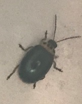
The beetle wing has a green, iridescent, sheen, unlike many others. My first thought was that the green must come from some particular element impurity present in the wings. However, a detailed imaging analysis using energy dispersive x-rays in the electron microscope was unable to capture anything besides carbon! This could either indicate that there is predominantly only carbon, and that the carbon is somehow organized structurally to give the green color. Or the impurity elements are present however not so easily identified using the method of analysis with energetic electrons. These electrons are often accelerated to greater than 1 keV energies, and so this is rather amazing to be able to see the organic matter magnified to the point where fine features not possible to see clearly with a basic optical microscope are visible and in focus in the electron microscope.
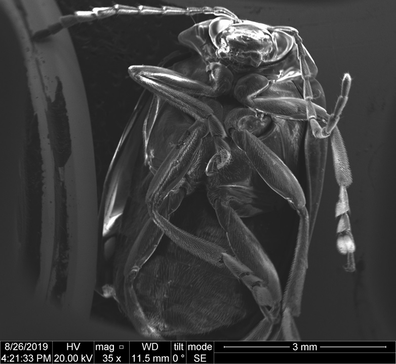
Looking closely at the beetle, several anatomical regions certainly pique the interest: the eyes, the face/mandibles(?), antennae and legs. While I am not an insect scientist, I can certainly enjoy looking at these images.
Part II. Dust Mites – (Not fully identified)
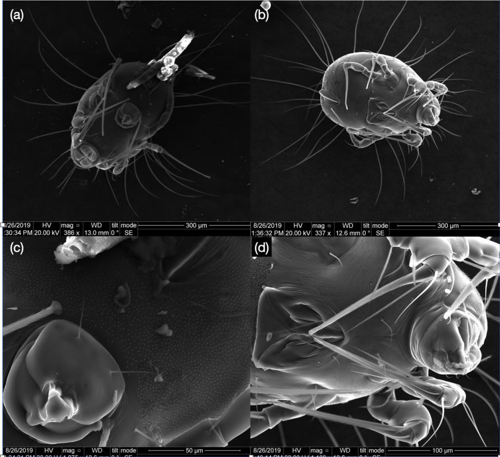
The species of mite imaged here may be similar to the Blomia tropicalis, a house dust mite found in the tropics regions. These mites, are tentatively identified as within the glycyphagidae family of arachnid class of phylum arthropods [1-3]. With eight legs and spindly hairs, these animals share many similarities with spiders, scorpions, harvestmen, amongst others. The identification of these mites compared with others may be possible from the images of the tarsi and other structures, Fig. 3. Compared to the Dermatophagoides pteronyssinus, these mites are different in size and shape [4].
Two distinct specimens are imaged in this report, possibly a female and male of the species. These identifications are made based on the genital structures below what should be identified as the thorax, Fig. 3(c, d). Both male and female contain numerous setae on their undersides, although the chaetotaxy (and function) is not yet clear.
Possible pathologies are respiratory disease and asthma, as well as dermatological abrasion (these mites cause a terrible itch! 🙂 ) [5]. Cysteine (only sulfur containing amino acid of the 26) proteases are identified in the allergens of B. tropicalis although protease activity in not fully determined experimentally. As this mite is within the arachnid family, the possible interactions in these processes are of interest for those into bugs. Possible allergen from blot studies with IgE suggest that specific structures (such as Blot 5, 6 and 13) contain higher probability for inducing immune response in humans.
I am not an expert on this, and for the time being, I plan on leaving my understanding of these fascinating organisms at the level of imaging. I found these two specimens fortuitously and placed them on a piece of carbon tape for the imaging in the scanning electron microscope. Rest assured, if I see one crawling on me, I plan on pinching the bug off me or taking a shower.
References:
- https://www.jw.org/en/publications/magazines/g201210/sensors-of-black-fire-beetle/
- “Glycyphagus”. Accessed on December 25, 2020. https://en.wikipedia.org/wiki/Glycyphagus.
- L. Guilleminault, C. Viala-Gastan. “Blomia tropicalis: un acarien sous les tropiques (Blomia tropicalis: A house dust mite in the tropics)”. Revue des Maladies Respiratoires Volume 34, Issue 8, October 2017, pp. 791-801. https://www.sciencedirect.com/science/article/abs/pii/ S0761842517300451.
- Gaud, Jean; Atyeo, Warren T. (1996). “Feather mites of the world (Acarina, Astigmata): the supraspecific taxa”. Annales-Musee Royal de l’Afrique Centrale. Sciences Zoologiques (Belgium). Musee Royal de l’Afrique Centrale. https://www.nhbs.com/feather-mites-of-the- world-acarina-astigmata-the-supraspecific-taxa-2-volume-set-book.
- “House dust mites”. Accessed on December 25, 2020. https://en.wikipedia.org/wiki/ House_dust_mite.
- Eduardo Santos da Silva, Claudia Asam, Peter Lackner, Heidi Hofer, Michael Wallner, Carina Silva Pinheiro, Neuza Maria Alcantara-Neves, Fatima Ferreira. “Allergens of Blomia tropicalis: An Overview of Recombinant Molecules”. Int. Arch. Allergy Immunol. 2017 Apr. 29; 172 (4): 203-214. https://www.ncbi.nlm.nih.gov/pmc/articles/PMC5472214/.